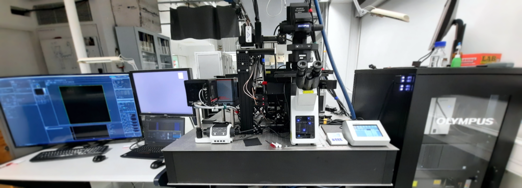
Fast Coherent Raman Imaging

Contribution of the University of Helsinki to the infrastructure includes construction of a fast coherent Raman imaging instrument at the Department of Chemistry. At present such instrument is not yet commercially available but has been demonstrated in leading analytical laboratories (e.g. at Harvard University, Purdue University) to have enormous potential in the material and life sciences to provide chemical and supramolecular insights not revealed using established state-of-the-art analytical methods.
qCSI Helsinki is now operated by LifeSpec HiLIFE infrastructure and is available for academic and commercial use. Follow the link for more information.
Fast coherent Raman imaging is a unique technology enabling label-free, global, quantitative, and non-destructive submicron 3D imaging of diverse and dynamic samples, in both the material and life sciences. The technique is based on vibrational spectroscopy via inelastic Raman scattering. Compared to the classical spontaneous Raman effect, the coherently driven transitions result in an enhancement of up to 105 in signal intensity. This overcomes the biggest analytical barrier with conventional state-of-the-art Raman mapping: painfully slow image acquisition times of up to hours or days. Furthermore, the spatial resolution is improved and one-photon fluorescence interference is also avoided (especially important for life sciences). Standard coherent Raman microscopes are very fast (video-rate imaging), but operate in narrowband spectral mode corresponding to a single vibrational transition. This severely limits the ability to resolve different chemical species and supra-molecular structures. The coherent Raman imaging instrument will instead allow a broad spectral coverage (currently 1100-3200 cm-1), enabling fast quantitative imaging of chemically and structurally complex systems. Inherently confocal non-contact sampling and the lack of water signal makes the technique ideal for 3D imaging in situ/in vitro/ex vivo of e.g. cells, tissues, microfluidics.
The main focus is in stimulated Raman scattering (SRS) with spectral focusing technique, which allows fast frame-by-frame measurement of vibrational spectral images. This is complemented by sum frequency or second harmonic generation (SFG/SHG) and two-photon excited fluorescence (TPEF), and normal confocal fluorescence imaging to further increase chemical and structural specificity. The non-destructive analyses also enable complementary correlative coupling with, e.g. mass spectrometry for metabolomics, proteomics, and reaction monitoring, and transmission and scanning electron microscopy for further structural analysis.
Further reading on coherent Raman imaging:
Evans, C. L., & Xie, X. S. (2008). Coherent anti-Stokes Raman scattering microscopy: chemical imaging for biology and medicine. Annual Review of Analytical Chemistry, 1, 883-909.
Cheng, J. X., & Xie, X. S. (2015). Vibrational spectroscopic imaging of living systems: An emerging platform for biology and medicine. Science, 350(6264).
Tipping, W. J., Lee, M., Serrels, A., Brunton, V. G., & Hulme, A. N. (2016). Stimulated Raman scattering microscopy: an emerging tool for drug discovery. Chemical Society Reviews, 45(8), 2075-2089.
Fu, D., Holtom, G., Freudiger, C., Zhang, X., & Xie, X. S. (2013). Hyperspectral imaging with stimulated Raman scattering by chirped femtosecond lasers. The Journal of Physical Chemistry B, 117(16), 4634-4640.
